
Figure 1.
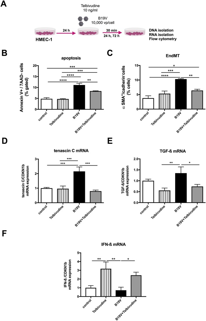
Impact of telbivudine on parvovirus B19‐infected human microvascular endothelial cells. (A) Experimental design: human microvascular endothelial cells (HMEC)‐1 were 24 h after plating, infected with 10 000 viral particles/cell with/out 10 ng/mL of telbivudine, and collected after 30 min for the analysis of tenascin‐C and TGF‐β1 mRNA expression, after 24 h for the analysis of apoptosis, and after 72 h for the anaylsis of endothelial‐to‐mesenchymal transition (EndMT). Bar graphs represent the mean ± SEM of (B) Annexin V/7AAD‐ cells (% gated), (C) α‐smooth muscle actin (SMA)+/VE‐cadherin‐ cells (% gated), (D) tenascin‐C, (E) transforming growth factor (TGF)‐β, and (F) interferon (IFN)‐β mRNA expression, as indicated, with n = 4–6/group and *P < 0.05, **P < 0.01, ***P < 0.001, and ****P < 0.0001.