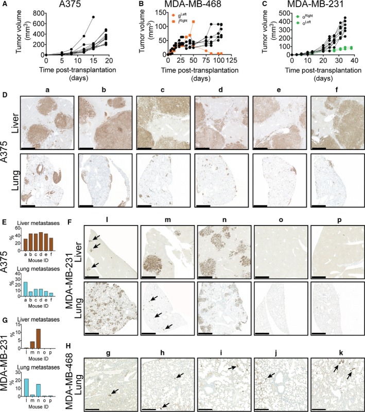
Figure 1.

Growth and metastasis formation in HIS mice. Tumors from the cell lines (A) A375 (n = 6 mice), (B) MDA‐MB‐468 (n = 5 mice), and (C) MDA‐MB‐231 (n = 5 mice) all expanded despite the presence of an allogeneic immune system. Primary tumors derived from A375 consistently yielded multiple large liver and lung metastases (D,E), which was only occasionally observed in mice carrying MDA‐MB‐231‐derived primary tumors (F,G). Few cancer cells were detected in the lungs of MDA‐MB‐468‐transplanted mice (image showing the most densely cancer cell‐containing areas) (H) and were absent in the liver. Cancer cells were detected by IHC staining using antibodies against human EGFR (A375) or pan‐cytokeratin (MDA‐MB‐231 and MDA‐MB‐468). Scale bar 1 mm (D,F) and 250 μm (H), respectively.
