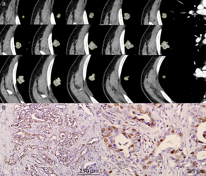
Figure 1.

Examples of segmentation of lung cancer based on contrast‐enhanced computed tomography images and Ki‐67 status. (a) Semiautomatic tumor segmentation was performed on each slice of the tumor using a three‐dimensional slicer, which showed a high Ki‐67 expression level (b, magnification ×100; c, ×400).
