
Figure 2. Sympathoexcitation by photostimulation of the nerve terminals of PVN‐RVLM neurons.
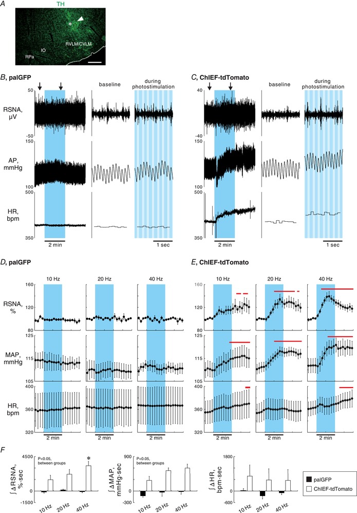
A, a medullary coronal section showing TH immunoreactivity and the scar (arrowhead) and wreck (asterisk) made due to an insertion of an optical fibre. Scale bar: 500 μm. The rostrocaudal level of this section was approximately 1 mm caudal to the caudal pole of the facial nucleus. CVLM, caudal ventrolateral medulla; RPa, raphe pallidus nucleus. B and C, representative traces of renal sympathetic nerve activity (RSNA), arterial pressure (AP) and heart rate (HR) while 2 min photostimulation at 40 Hz with 5 ms pulse duration was unilaterally given to the RVLM ipsilateral to the side of AAV injection into the PVN (blue background), obtained from palGFP (B) and ChIEF‐tdTomato (C) rats. Traces at baseline and during photostimulation (arrows in the left panels) are magnified in the right panels. bpm, beats per minutes. D and E, 15 s‐averaged time courses of RSNA, mean AP (MAP) and HR in 4 palGFP (D) and 8 ChIEF‐tdTomato (E) rats while photostimulation was given to the RVLM for 2 min at 10 Hz with 50 ms pulse duration (PD), 20 Hz with 10 ms PD or 40 Hz with 5 ms PD. Values are means ± SEM. Horizontal red bars, P < 0.05 vs. baseline. F, comparisons between the rat groups and among the photostimulation frequencies of RSNA, MAP and HR responses to photostimulation of the RVLM, assessed by integration of changes from baseline during 2 min photostimulation. * P < 0.05 vs. 10 Hz.