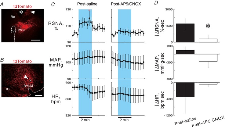
Figure 4. Effect of blockade of ionotropic glutamate receptors in the RVLM on PVN photostimulation‐elicited sympathoexcitation.

A, a hypothalamic coronal section showing a scar (arrowhead) and a wreck (asterisk) due to chronic implantation of an optical fibre directed to the PVN and tdTomato‐positive signals (autofluorescence) in a ChIEF‐tdTomato rat. Re, nucleus reuniens in the thalamus. Scale bar: 500 μm. B, a medullary coronal section of a ChIEF‐tdTomato rat, showing a scar (arrowhead) due to repetitive insertions of the pipette for microinjection in the RVLM, in which PVN‐derived, tdTomato‐immunoreactive axonal signals were concentrated. Scale bar: 500 μm. C, 15 s‐averaged time courses of RSNA, MAP and HR in six ChIEF‐tdTomato rats while 2 min photostimulation at 40 Hz with 5 ms pulse duration was given to the PVN 10–15 min after saline or a cocktail of AP5 and CNQX was injected into the RVLM. Horizontal red bar, P < 0.05 vs. baseline. D, comparisons of RSNA, MAP and HR changes in response to photostimulation of the PVN after injection into the RVLM with saline or a cocktail of AP5 and CNQX. * P < 0.05 vs. post‐saline.
