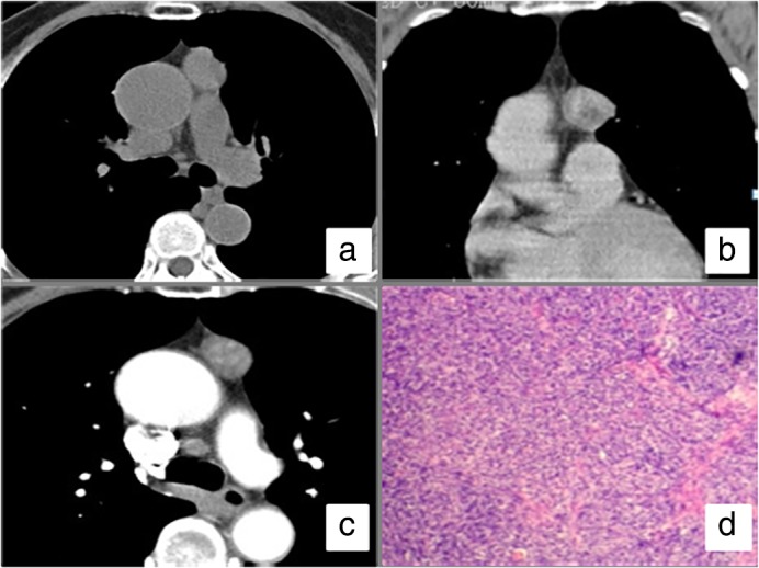
Figure 2.

Computed tomography (CT) scan of a thymoma. (a) A plain CT scan of the chest demonstrated an irregular node located in the anterior mediastinum; (b) coronal view of the chest demonstrated an irregular node next to the aorta; (c) an enhanced CT scan showed the node was significantly enhanced compared to the plain CT scan; (d) post surgery pathology showed thymoma.
