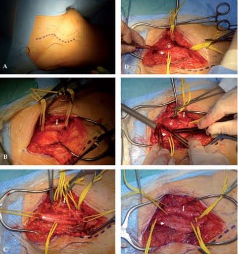
Figure 2.

Case 1. Intraoperative phases of surgical removal of the synovial cyst. A) patient in supine position. The longitudinal surgical approach has been drawn on the cutis. B) Surgical isolation of the femoral artery (white arrow) that appeared dislocated superficially. C) Surgical isolation of the femoral nerve (white head-arrow) that also appeared to be compressed by the mass. D) The synovial cyst (asterisk) has been detected deeper to femoral nerve and femoral vessels. E) Marginal excision of the synovial cyst through the cystic wall up to the joint capsule. F) Surgical field after excision of the synovial cyst
