Figure 3.

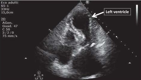
Echocardiogram recorded while in the ED, showing the typical apical ballooning, or takotsubo-like shape, of the left ventricle

Echocardiogram recorded while in the ED, showing the typical apical ballooning, or takotsubo-like shape, of the left ventricle