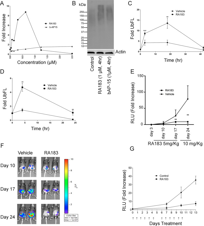
Figure 6.
RA183 inhibits proteasomal activity and tumor growth in mice. (A, B) 293TT cells were stably transfected with 4UbFL expressing plasmid35 and treated with titrations of the indicated compounds for 4 h. Lysates were prepared and luciferase activity was measured (A). Data are presented as fold change in luciferase activity compared to untreated cells. (B) Levels of Lys48-linked ubiquitin in lysates were analyzed by Western blot. (C) Mice were administered 4UbFL DNA to the skin via gene gun (i.d.). After 24 h, the site of DNA administration was treated topically by application of 4% (w/v) RA183 (40 μL) in emu oil or oil alone (10 mice/group) respectively. After the indicated time points post-treatment, bioluminescence was measured by intraperitoneal (i.p.) injection of luciferin and imaging with an IVIS 200 at the site of DNA delivery (* p <0.05, ** p <0.01). (D) Mice were administered 4UbFL DNA to the leg muscle (intramuscularly (i.m.)) via in vivo electroporation. RA183 was given i.p. at 20 mg/kg. After the indicated time points post-treatment, bioluminescence was measured in control and RA183-treated animals (10 mice/group) by intraperitoneal injection of luciferin and imaging with an IVIS 200 at the site of DNA delivery. (E, F) 3 × 10⁵ ID8-Luc cells (a syngeneic mouse ovarian cell line expressing luciferase) were injected into the peritoneal cavity of C57BL6 mice (five per group) on day 0. The mice were initially treated daily i.p. with either vehicle or 5 mg/kg RA183 dissolved in 2% DMSO and 25% β-hydroxypropyl cyclodextrin on days 3–17 and then RA183 dose was escalated to 10 mg/kg on days 17–24. The tumor growth was detected by imaging the mice through IVIS system and measuring the luciferase activity (relative light units (RLUs)) before, at the interval, and after the treatment. (G) Nude mice were administered ES2-luc cells i.p., and 2 days later, a basal bioluminescent level was determined for each mouse. Nude mice bearing ES2-luciferase cells i.p. were treated with either 6.6 mg/kg RA183 (dissolved in 2% DMSO and 25% β-hydroxypropyl cyclodextrin) (i.p.) or vehicle alone (n = 9 per group) 6 days on and 1 day off. Prior to and both 7 and 14 days after initiation of treatment, the mice were imaged for their luciferase activity (RLU) and fold change over baseline was determined.

