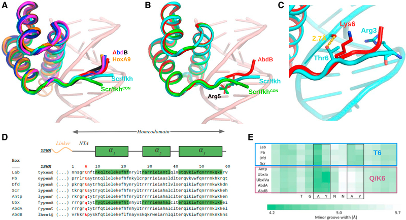
Figure 2. Distinct NTA Conformations of Anterior and Posterior Hox Proteins.
(A)Comparison of NTA conformations seen in four AbdB-Exd complexes and two Scr-Exd complexes.
(B)Same as (A) but showing only AbdB-Exd (red) and both Scr NTAs, highlighting the shared insertion of Arg5, which inserts into the minor groove at the same position in all of these Hox-Exd complexes.
(C)Close-up of AbdB-Exd (red) and Scr-Exd (fkh) NTAs showing that Thr6 of Scr, but not Lys6 of AbdB, makes a hydrogen bond with the phosphate backbone.
(D)Sequence alignment of Hox W-motifs and homeodomains, with position 6 highlighted in red.
(E) Data from Slattery et al. (2011), showing the selection of sequences with two minor groove width minima for anterior Hox proteins (Lab, Pb, Dfd, Scr) and only a single minor groove width minimum for posterior Hox proteins (Antp, Ubx, AbdA, AbdB). All Hox proteins preferring a second minimum have a threonine at position 6 (see D).

