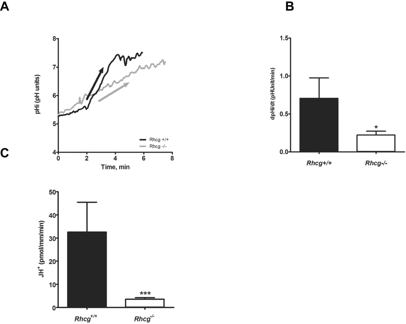
FIGURE 2. H+ secretion is reduced in cortical collecting duct cells from Rhcg−/− mice.
Cortical collecting ducts were isolated from kidneys of Rhcg+/+ and Rhcg−/− mice after 2 days of HCl-loading, microperfused in vitro, and intracellular pH (pHi) monitored. NH4Cl (20 mM) was applied from the luminal perfusate. (A) Original pHi tracing from a CCD exposed to a luminal NH4Cl pulse. The arrow indicates the rate of pHi change measured and calculated. Exposure to an NH4Cl pulse caused a large intracellular acidification (not shown) followed by an alkalinization phase leading to pHi recovery. The initial slope (ΔpHi/Δt) of the alkalinization phase was measured . (B) Bar graph summarizing ΔpHi/Δt after removal of the luminal NH4Cl pulse (n = 8–12 tubules/genotype). (C) Bar graph summarizing H+-fluxes calculated based on intracellular buffering power and pHi recovery rates.

