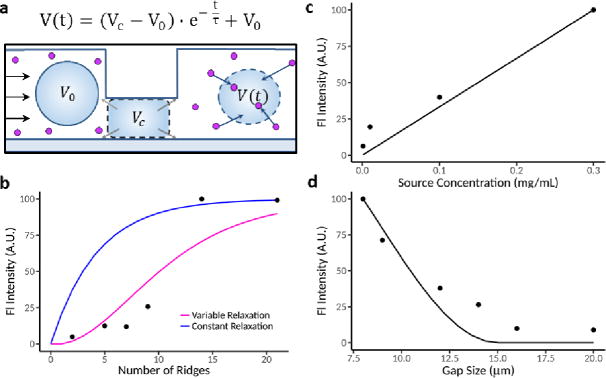
Figure 4.

Development of mechanistic model to incorporate cell volume exchange. (a) An illustration showing the assumptions made in our model. Before encountering any ridges, cells have initial volume V0. Under each ridge, we assumed cell volume decreased to some constant VC which was dependent on the ridge gap. After clearing the ridge, we assumed that the cell volume increases with time t after ridge compression and approaches V0 asymptotically with timescale τ. Cell relaxation was assumed to either be constant or to occur more rapidly with more compressions. Total volume exchange is therefore dictated by the amount that the cell relaxes between ridges. (b-d) Comparisons between the median fluorescence intensity observed in our experiments (dots) to the predictions of our model (solid lines).
