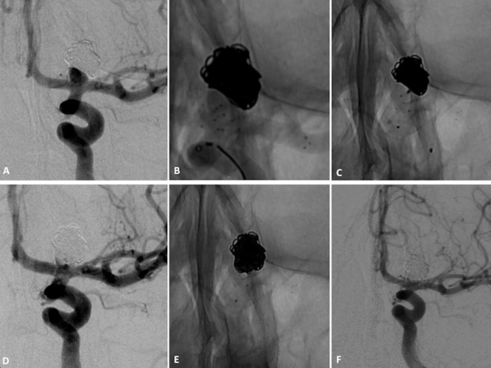
Figure 3.
Patient with left unruptured internal carotid artery bifurcation aneurysm. (A) Subtracted angiogram of internal carotid artery shows a wide-necked terminus carotid aneurysm. (B) Non-subtracted image shows Barrel vascular reconstruction device (VRD). The six markers of the cage covering aneurysmal neck are shown. (C) Non-subtracted image shows the placement of a second microcatheter through struts into the aneurysm fundus. (D) Non-subtracted image at the end of coiling. (E) Subtracted angiograms of the internal carotid artery at the end of the procedure show small neck remnant and one in-stent thromboembolic event. (F) Subtracted angiogram of internal carotid artery at 12-month follow-up shows near-complete occlusion with neck remnant that is stable in size.

