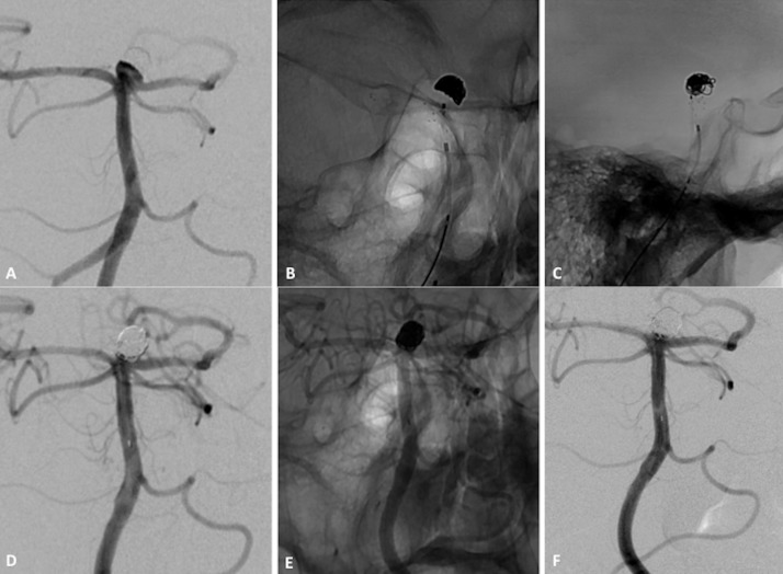
Figure 4.
Patient with wide-necked basilar tip aneurysm. (A) Subtracted angiogram of left vertebral artery shows small basilar tip aneurysm with 4 mm neck. (B, C) Non-subtracted image after placement of Barrel vascular reconstruction device (VRD). (D, E) Subtracted and non-subtracted angiograms of vertebral artery at procedure end show complete obliteration of neck remnant. (F) Angiogram at 12-month follow-up shows complete aneurysm occlusion.

