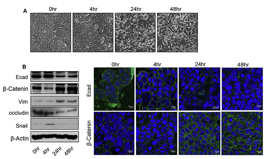
Fig. 1.
TNFα induces epithelial to mesenchymal transition (EMT) in human colon adenocarcinoma HT29 cells. A. Representative phase-contrast images of HT29 cells growing in monolayer cultures. Treatment of TNFα (10 ng/ml) induced morphological alterations characterized as fibroblast-like cells with significant changes at 24 and 48 h. B. Immunoblot analysis of HT29 cells treated with TNFα for 0–48 h. Cells were lysed in RIPA Buffer, and a total of 20 μg proteins for each sample were loaded onto the SDS-polyacrylamide gel. Membranes were blotted against E-cadherin, β –catenin, vimentin, and occludin whereas actin was used as a loading control. C. Immunofluorescence illustration for localization of E-cadherin and β-catenin with DAPI in HT29 cells treated with TNFα for time dependent manner (0–48 h).

