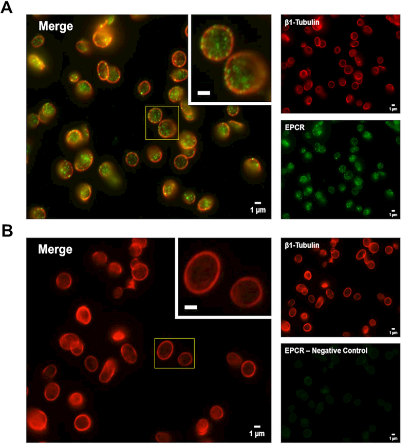
Figure 7. Human platelets contain pre-formed EPCR.

(A and B) Freshly isolated human platelets (unactivated) were fixed in 4% formaldehyde and spun down onto poly-L-lysine-coated glass coverslips. Samples were permeabilized with 0.5% Triton-X-100 and incubated overnight in blocking buffer prior to probing for EPCR and β1-tubulin using appropriate primary and secondary antibody pairs conjugated to Alexa Fluor 488 or 568. Samples were examined with a Nikon TE-2000-E Microscope (Nikon, Tokyo, Japan) equipped with a 100x (N.A. = 0.3) Plan-Fluor objective. Images were obtained using a charged coupled device (CCD) camera (Hamamatsu Photonics, Bridgewater, NJ). Background fluorescence was subtracted and image brightness/contrast was adjusted linearly for each micrograph. Larger panels (left) represent a merged image of the two smaller panels (right) for each image. Insets represent a magnified region outlined by the yellow box for each image (inset bars = 1μm). (A) β1-tubulin (red) was used to label the platelet cytoskeleton while EPCR (green) is predominantly localized in distinct, punctate granules within the platelets. (B) Non-specific staining was assessed by labeling in the absence of the primary anti-EPCR antibody (green secondary antibody alone).
