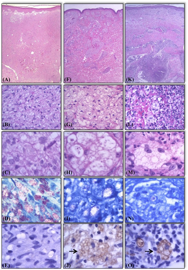
Figure 2.
Histological and bacilloscopic characteristics of lesions of lepromatous side in activity, lepromatous side in regression, and R2 lesions. Active lesions with vacuolated macrophages containing a large number of solid bacilli (A–D). Lesions in regression showing multivacuoled macrophages containing a large number of multifragmented bacilli (F–I). R2 lesions, the macrophages display characteristics similar to those seen in lesions in regression undergoing an influx of neutrophils (K–N). Expression of AKR1B10 in the same samples. Negative expression in samples of lepromatous side in activity (E). Positive expression (marked by →) in samples of lepromatous side lesions in regression (J), and R2 lesions (O). Hematoxylin-eosin staining (A–C, F–H, K–M). Fite-Faraco staining for bacilloscopy (D,I,N).

