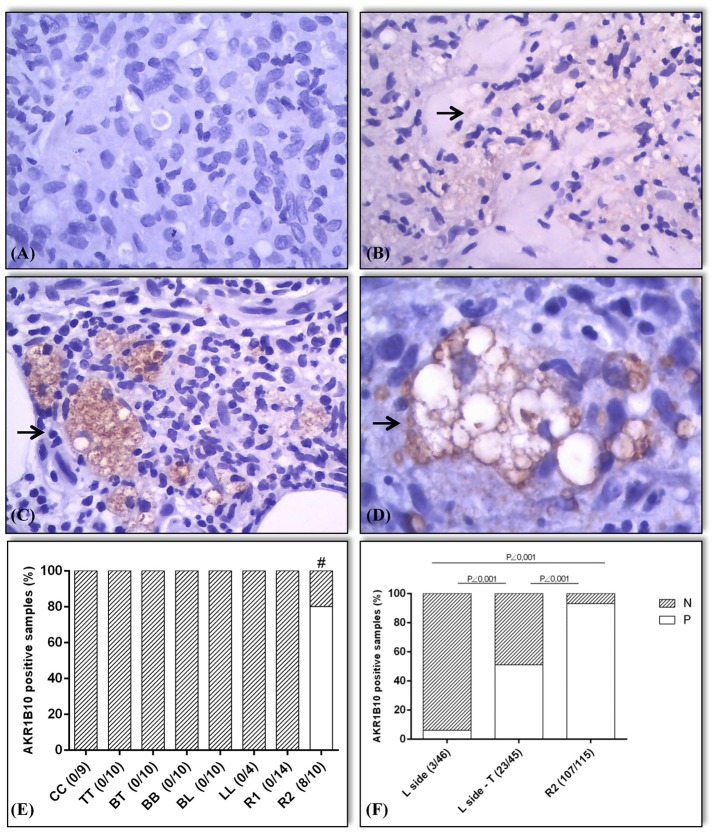
Figure 3.
Expression pattern (marked by →) and expression values of AKR1B10 in positive samples. The samples were classified as negative for AKR1B10 when its expression was absent (0) or weak (1+; A,B). Positive expression of AKR1B10 (2+ or 3+ on the scale; C,D). In the preliminary assessment (E), immunostaining was negative in all samples of HC, TT, BT, BB, BL, LL, and R1. It was positive in 8 out of 10 samples of R2 (80%), with a significant difference between groups (# R2 vs. HC, R2 vs. TT, R2 vs. BT, R2 vs. BB, and R2 vs. BL, all with p = 0.0007; R2 vs. LL, p = 0.015; R1 vs. R2, p < 0.0001). In later evaluation (F), there was positive expression in 3 out of 46 active lepromatous (BL and LL) samples (6%) (L side), 23 out of 45 samples of BL + LL in regression after treatment (L side-T) (51%), and 107 out of 115 samples of R2 (93%), with a significant difference between the L side samples and L side-T (p < 0.0001), R2 and L side (p < 0.0001), and between R2 and L side-T groups (p < 0.0001).

