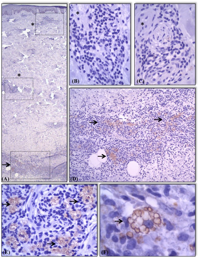
Figure 4.
Expression of AKR1B10 in different components of skin and granulomas in the samples. Absence (marked with a*) of AKR1B10 expression in most components of skin and in some granulomas (A–C). Positive expression (marked with a →) in granulomas present in other areas of the same sample (A,D). Moderate or strong expression of marker almost exclusively in the cytoplasm of macrophages present in lesions of lepromatous lesions in regression (E) and R2 (F) showing staining of intracytoplasmatic vacuoles/lysosomes (F).

