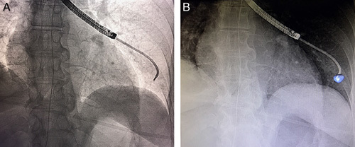
FIGURE 2.

Comparison of standard fluoroscopy (A) and augmented fluoroscopy (B) for a fluoroscopically invisible nodule. The blue volume was segmented from cone-beam computed tomography data and automatically projected using dedicated software (OncoSuite; Philips).
