
Figure 6.
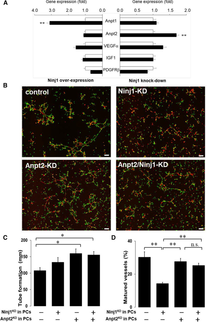
Role of angiopoietin in Ninj1 (ninjurin1)–mediated vascular formation. A, Expression of angiogenesis-related genes in primary cultured pericytes (PCs). Ninj1 expression was increased (overexpression) or decreased (knockdown) by transfection with Ninj1-expressiong vectors or Ninj1-specific small interfering RNA (siRNA), respectively (black bars), and their controls (white bars). After 2 d incubation, indicated gene expression was determined by quantitative polymerase chain reaction. Changes in mRNA after Ninj1 overexpression or knockdown were calculated. Mean±SEM (n=6, **P<0.01 vs white bar, t test analysis). B, After transfection of Ninj1- and Anpt (angiopoietin) 2-siRNA (Ninj1 knockdown [KD], Anpt2 KD, respectively) or their scramble-siRNA (control), DsRed-endothelial cells (red) and primary PCs isolated from GFP transgenic mice (green) were mixed and incubated in Matrigel. Fluorescence images are shown. C, The total lengths of formed tubes in the gels were measured. D, The ratio of matured vessels to total length of formed tubes is demonstrated. Mean±SEM (n=6, *P<0.05, **P<0.01, n.s. indicates not significant, ANOVA).