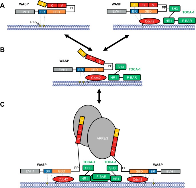
Figure 9.
Model for WASP activation. WASP is represented as in Fig. 1, together with binding partners PIP2 (labeled, yellow circle), Cdc42 (red oval), TOCA1 (green, composed of domains SH3, HR1, and F-BAR), and Arp2/3 (gray ovals). The cell membrane is represented by a blue bilayer with which PIP2 and Cdc42 are associated. A, WASP binding to PIP2 (left panel) or WASP binding to Cdc42 (right panel) does not stimulate VCA release alone, but binding of both can relieve autoinhibitory interactions to release the VCA region. B, this can then bind to Arp2/3, which is fully activated by binding two VCA regions. C, dimerization through scaffolds such as TOCA1 can facilitate Arp2/3 activation by two VCA regions.

