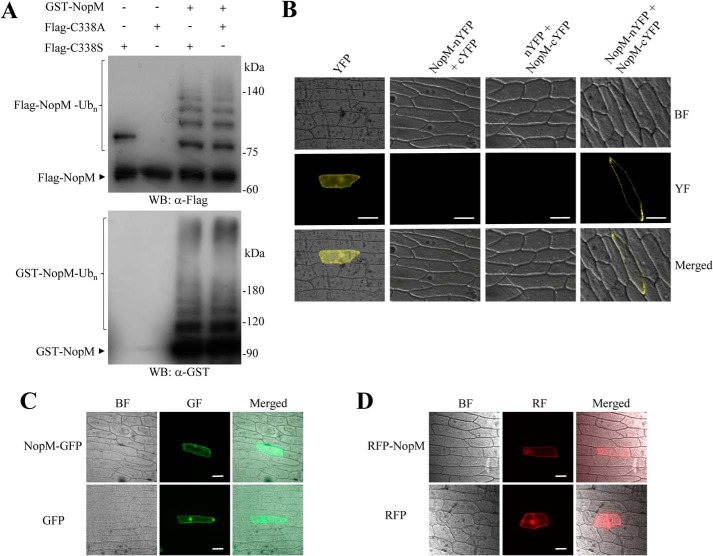
Figure 3.
Analysis of NopM–NopM interactions and subcellular localization of NopM fused to fluorescent proteins. A, analysis of intermolecular transfer of ubiquitin in vitro. The Flag-tagged NopM variants C338A (enzymatically inactive) and C338S (forming a mono-ubiquitinated conjugate) were ubiquitinated by GST–NopM. Western blot (WB) were probed with anti-Flag or anti-GST antibodies. B, BiFC analysis of NopM–NopM interactions in vivo. Onion cells expressing indicated protein combinations were microscopically analyzed for yellow fluorescence (YF) emission and under bright field (BF) illumination. Co-expression of NopM–nYFP with NopM–cYFP resulted in formation of a BiFC complex at plasma membranes. Bars, 100 μm. C and D, subcellular localization of full-length NopM fusion proteins. Fluorescent NopM proteins with C-terminal GFP tag (C) or with N-terminal RFP tag (D) were expressed in onion cells. GFP and RFP alone were expressed for comparison. Emission of green fluorescence (GF), red fluorescence (RF), and bright field conditions were used for microscopic examination. Bars, 100 μm.

