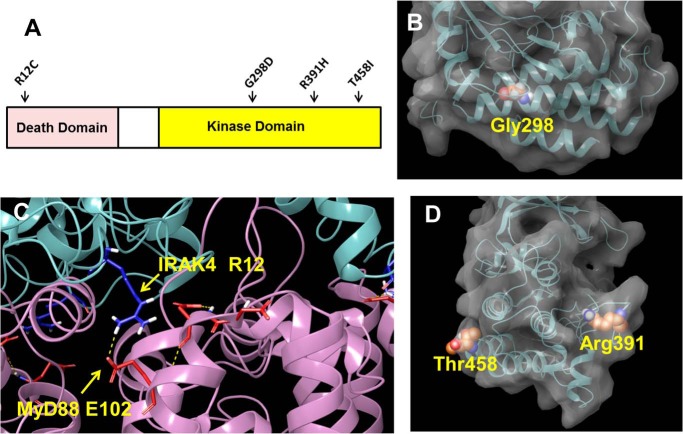
Figure 3.
Position of mutations in IRAK4 structure. A, cartoon showing position of human mutations in IRAK4. B, space-fill representation of Gly-298 in the kinase domain of IRAK4 taken from PDB entry 2NRU. C, interaction of IRAK4 Arg-12 with Glu-102 of MyD88 taken from PDB entry 3MOP. D, space-fill representations of Thr-458 and Arg-391 in the kinase domain of IRAK4 taken from PDB entry 2NRU. Space-fill representations were created using the Maestro software program (Schrödinger LLC, New York).

