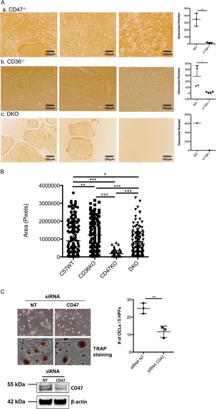
Figure 2.
CD47 is not required for osteoclastogenesis in vitro. A, panel a shows representative images from cocultures of osteoblasts isolated from neonatal C57WT mice and bone marrow cells isolated from CD47−/− mice. The first two images are from cocultures treated with a control IgG antibody. Osteoclasts are present in both images, although in some areas the osteoclasts are small. The right-most image in panel a is from a coculture treated with the neutralizing antibody to TSP-1. The scatter plot to the right of panel a summarizes the effect of TSP-1 antibody on osteoclastogenesis in three cocultures using CD47−/− preosteoclasts (n = 3; **, p ≤ 0.003). In the absence of TSP-1 antibody, the number of osteoclasts formed was equivalent to that seen in WT mice when CD47−/− marrow was used as the source of osteoclast precursors. Panel b shows representative images from cocultures of osteoblasts isolated from neonatal C57/BL/6j mice and bone marrow cells isolated from CD36−/− mice. Again, the first two images are from cocultures treated with a control IgG antibody. Abundant osteoclasts formed, and this effect was completely blocked by neutralizing TSP-1 (right-most image in panel b). The scatter plot to the right in panel b summarizes the effects of the neutralizing antibody on osteoclast formation (n = 4; *, p ≤ 0.02). Panel c shows representative images from cocultures of osteoblasts isolated from neonatal C57/BL/6j mice and bone marrow cells isolated from mice in which the genes for both CD47 and CD36 were deleted (DKO). Osteoclast formation occurred when control IgG was added in the first two images but was completely blocked by anti-TSP-1 antibody (third image and scatter plot to the right). Panel c represents data from one pair of cocultures. Consequently, no formal statistical analyses of data in panel c were performed. B, mean area of mature osteoclasts differentiated from WT osteoclast precursors (C57/BL/6j; left column), osteoclast precursors isolated from CD36−/− mice (second column from the left), osteoclast precursors isolated from CD47−/− mice (third column from the left), and osteoclast precursors isolated from DKO mice (far right column). Data are derived from 200 osteoclasts (C57WT versus CD36−/−; **, p ≤ 0.0017; for C57WT versus CD47−/−, for CD47−/− versus CD36−/−, for CD36−/− versus DKO, and for CD47−/− versus DKO, *** indicates p < 0.0001; for C57WT versus DKO, * indicates p ≤ 0.0490). C, human osteoclast-like cells generated from peripheral monocytes were treated either with a scrambled RNA (NT) or an siRNA directed against CD47. Suppressing CD47 did not abrogate osteoclastogenesis (upper left), although the number of osteoclasts formed was reduced by roughly 50% (upper right) (n = 3; **, p ≤ 0.0063). The scatter plots represent mean ± S.D. (error bars). Lower left, Western blot analysis demonstrates that the siRNA suppressed CD47 expression by 60–70%. CD47 has an apparent molecular mass on SDS-PAGE of ∼55 kDa. In the upper left image, the top panel is a phase-contrast image, and the bottom panel is from a TRAP-stained culture. HPFs, high-power fields.

