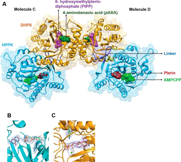
Figure 2.
Crystal structure of PvHPPK–DHPS. A, dimeric PvHPPK–DHPS where bound substrate/analogs in both HPPK and DHPS domains are shown as molecular surfaces. HPPK (cyan), DHPS (yellow), and the linker regions (blue) are marked. The bound substrates/analogs of pterin (maroon), AMPCPP (light green), PtPP (purple), pABA (green), and Mg2+ ion (lime green) are shown as sticks and spheres. B and C, the simulated annealing composite omit map contoured at 2σ levels for the bound ligands where ligands and bound Mg2+ ion are shown as sticks and spheres, respectively.

