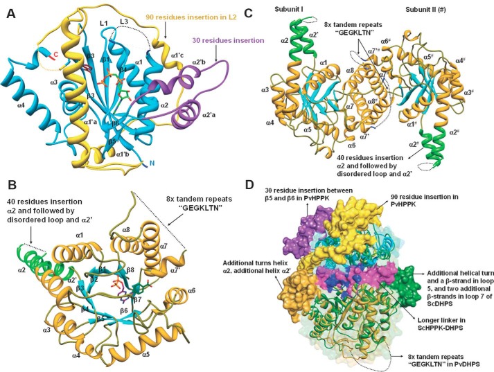
Figure 4.
PvHPPK–DHPS domains and structural comparisons. A and B, the domains of PvHPPK–DHPS are shown with secondary structural elements. The unique features/larger insertions are also labeled. C, a view of dimer interface and the participating helices α6, α7′, α7, and α8. A possible tandem repeat motif is shown. D, the superposition of DHPS domain of PvHPPK–DHPS on ScHPPK–DHPS where their unique features are highlighted.

