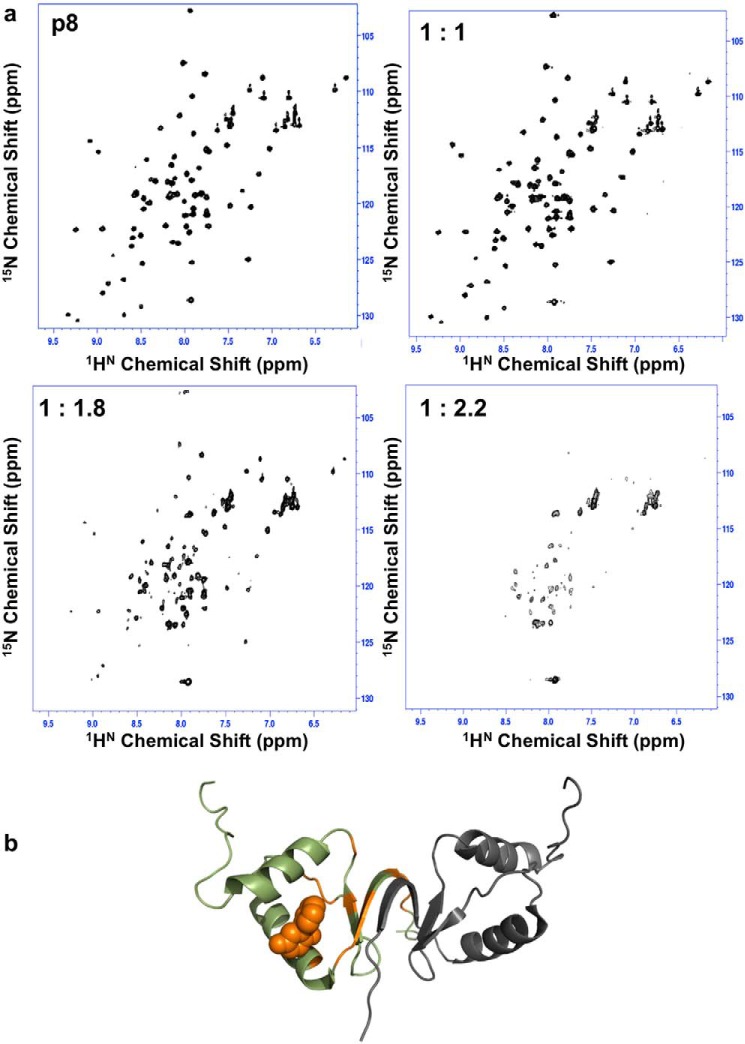
Figure 5.
Partial unfolding of p8 in the presence of compound 19. a, 1H-15N HSQC spectra recorded for p8 in the presence of increasing concentrations of compound 19, revealing gradual alteration of the quality of NMR spectra until partial protein denaturation for protein/ligand ratios exceeding 1:1.8. b, cartoon representation of the homodimer structure of p8 mapping positions of residues exhibiting severe line-broadening until the limit of NMR detection (orange) in the presence of compound 19, shown as an orange sphere (predicted binding pose obtained from docking).

