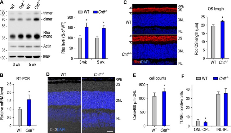
Figure 1.
Increase in rhodopsin (Rho) expression, thickness of the ONL, and length of rod OS in Cntf−/− mouse. A, immunoblot analysis of Rho and IRBP in 3- and 5-week-old WT and Cntf−/− retinas. Actin was detected to normalize sample loading. The histogram shows percentages of the immunoblot intensities of Rho from Cntf−/− retinas relative to the Rho immunoblot intensities of WT retinas. Asterisks in this figure indicate significant differences between WT and Cntf−/− mice (p ≤ 0.05); error bars indicate S.D. (n ≥ 3). B, transcription level of Rho mRNA in 3-week-old Cntf−/− retina was determined by quantitative RT-PCR and expressed as fold of Rho mRNA in age-matched WT retina. C, immunohistochemistry showing localization of Rho (red) to the rod OS in the superior retinas of 3-week-old WT and Cntf−/− mice. Nuclei were stained with DAPI (blue). Histogram shows Rho-positive OS lengths in the sections. D, differential interference contrast (DIC) images of retinal sections from 3-week-old WT and Cntf−/− mice. All scale bars denote 20 μm. E, rod nuclear numbers counted in a 400-μm wide superior ONL area that is 600 μm away from the optic nerve head. F, TUNEL-positive cell numbers in the ONL-OPL and inner nuclear layer (INL)-inner plexiform layer (IPL) of retinal sections from WT and Cntf−/− mice at postnatal day 8.

