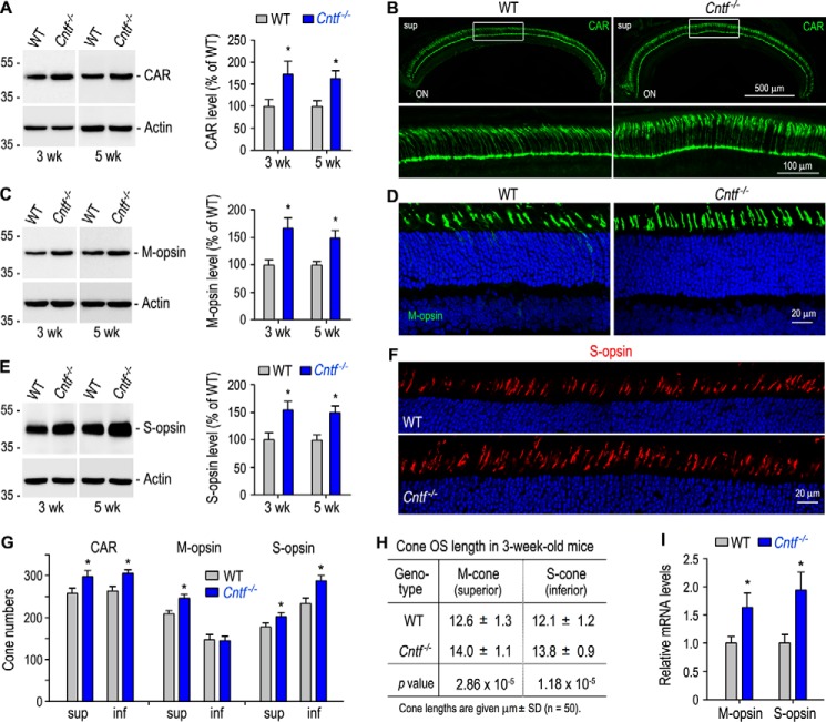
Figure 2.
Increase in expression of cone-specific proteins and length of cone OS in Cntf−/− mouse. A, immunoblot analysis of CAR in the retinas of 3- or 5-week-old WT and Cntf−/− mice. The histogram shows percentages of immunoblot intensities of CAR from Cntf−/− retinas relative to the CAR immunoblot intensities of WT retinas. B, immunohistochemistry showing CAR immunoreactivity in the superior (sup) retinas of 3-week-old WT and Cntf−/− mice (upper panels). ON, optic nerve. The areas of the rectangles are shown in the higher magnification images (bottom panels). C and E, immunoblot analysis of M-opsin (C) and S-opsin (E) from 3- or 5-week-old WT and Cntf−/− retinas. Histograms show percentages of immunoblot intensities of M-opsin (C) or S-opsin (E) in the Cntf−/− retinas relative to M-opsin or S-opsin intensities in WT retinas. D and F, immunohistochemistry for M-opsin (D) and S-opsin (F) in the superior (D) or inferior (F) retinas of 3-week-old WT and Cntf−/− mice. G, numbers of CAR- or cone opsin-positive cells in the superior (sup) or inferior (inf) retinas of 3-week-old WT and Cntf−/− mouse retinal sections taken from the dorsal-ventral midline of the eye. H, lengths of M- and S-cone OS in 3-week-old WT and Cntf−/− mouse retinal sections. I, relative mRNA levels of cone opsins in 3-week-old WT and Cntf−/− retinas were determined by quantitative RT-PCR. All asterisks indicate significant differences between WT and Cntf−/− mice (p < 0.05); error bars denote S.D. (n = 3∼4).

