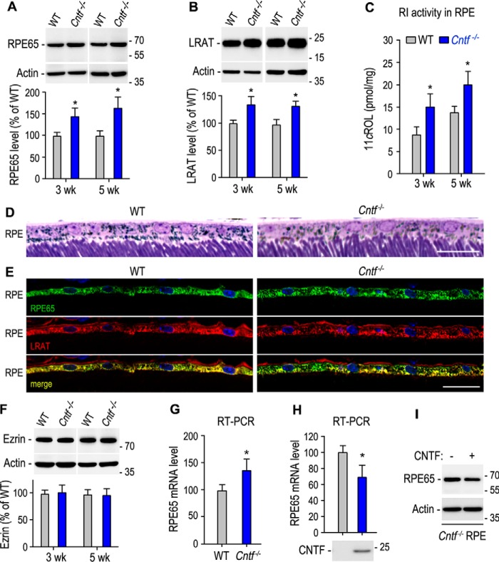
Figure 3.
Up-regulation of the visual cycle enzymes in the Cntf−/− RPE. A and B, immunoblot analysis of RPE65 (A) and LRAT (B) in the RPE of 3- or 5-week-old WT and Cntf−/− mice. Histograms show immunoblot intensities of RPE65 or LRAT from the Cntf−/− RPE relative to RPE65 or LRAT intensities in WT mouse RPE. C, retinoid isomerase (RI) activities determined by measuring synthesis of 11cROL from all-trans-retinol substrate incubated with RPE homogenates of 3- or 5-week-old WT and Cntf−/− mice. D, light microscopic images of RPE layers in the superior retinal sections of 5-week-old WT and Cntf−/− mice. E, immunohistochemistry showing distribution patterns of RPE65 and LRAT in the superior retinal sections from 3-week-old WT or Cntf−/− mice. Scale bars in D and E denote 20 μm. F, immunoblot analysis of Ezrin in the eyecups of 3- or 5-week-old WT and Cntf−/− mice. The histogram shows relative immunoblot intensities of Ezrin in WT and Cntf−/− eyecups. G, relative expression levels of RPE65 mRNA in WT and Cntf−/− RPE were determined by quantitative RT-PCR. H, quantitative RT-PCR showing relative expression levels of RPE65 mRNA in Cntf−/− mouse eyecup RPE incubated with media with or without CNTF. I, immunoblot analysis of RPE65 in Cntf−/− mouse eyecup RPE in H. All asterisks indicate significant differences between WT and Cntf−/− mice or between test and control groups (p < 0.04); error bars are S.D. (n = 3 or 4).

