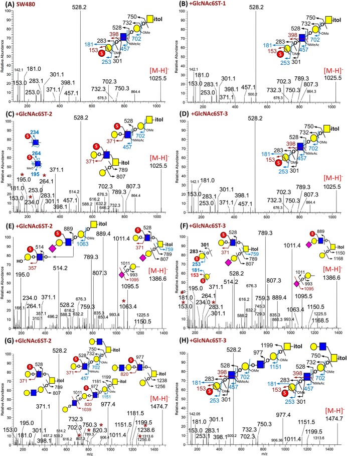
Figure 3.
Negative ion mode nanoESI-MS/MS analyses of permethylated monosulfated O-glycans from nontransfected and different GlcNAc6ST-transfected SW480 cells. A complete set of HCD MS2 spectra for eight major monosulfated O-glycans detected in all four cell lines is collated as Fig. S4. Only those for the precursors at m/z 1025 (A–D), 1386 (E and F), and 1474 (G and H) are shown here. A minimum number of possible isomeric structures are provided on each spectrum as cartoon drawings, with the origins of assigned fragment ions indicated. For nonsialylated O-glycans at m/z 1025, the diagnostic ions for GlcNAc6S were only detected in the spectrum of +GlcNAc6ST-2 cells (C). The sialylated structures at m/z 1386 carried exclusively GlcNAc6S in the +GlcNAc6ST-2 cells (E), but the additional presence of Gal3S-terminated type 1 chain was evident by the diagnostic ions for the corresponding structures made by the +GlcNAc6ST-3 cells (F). Signals at m/z 789/807 and 1238/1256 in G identified the additional presence of isomeric core 2 structures carrying 6-sulfo LacNAc on the 6-arm in the +GlcNAc6ST-2 cells, which were absent from the +GlcNAc6ST-3 cells (H). In each case, the critical ions indicative of isomeric structure assignment were marked with an asterisk.

