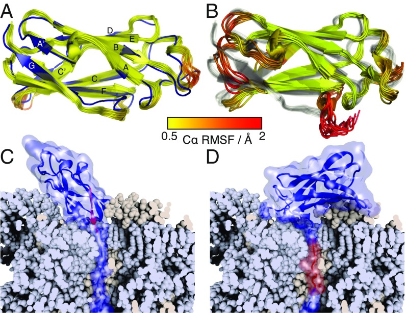
Fig. 2.
Structural analysis of FLN5 truncation variants. (A) Ensemble structure of FL FLN5 (yellow to red) aligned against the previously determined crystal structure (blue) (18). The FL ensemble is colored according to the C RMSF as indicated by the key. (B) Ensemble structure of the 6 trans intermediate (colored by the C RMSF) aligned against the FL ensemble (gray). (C and D) Modeling of the closest possible approach of (C) native and (D) intermediate FLN5 structures tethered to the ribosome (shown in a cutaway view to highlight the NC path through the exit tunnel). The G strand of FLN5 (disordered in the intermediate) is highlighted in red.

