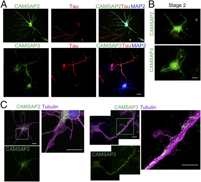
Fig. 1.
Distribution of CAMSAP2 and CAMSAP3 in hippocampal neurons in culture. (A) Neurons were triple-immunostained for CAMSAP2 or CAMSAP3 (green), Tau (red), and MAP2 (blue) at DIV6. (B) Neurons (stage 2) were immunostained for CAMSAP2 or CAMSAP3 at DIV2. (C) Neurons (stage 3) were triple-stained for CAMSAP2 or CAMSAP3, α-tubulin (magenta) and DNA (blue) at DIV4. Boxed areas are enlarged. (Scale bars, 20 μm in A and 10 μm in B and C.)

