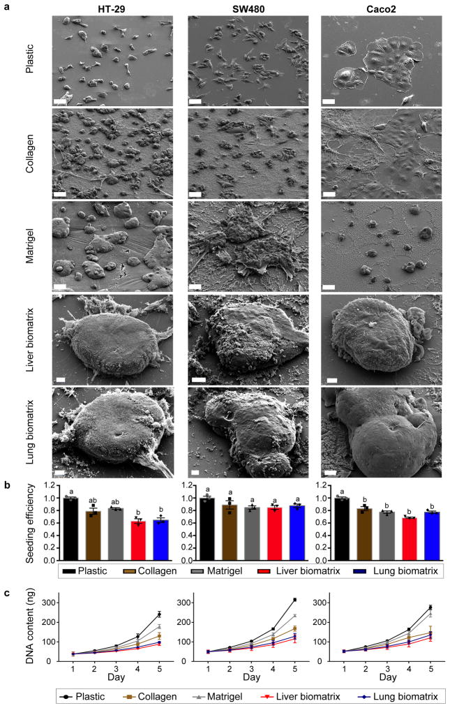
Figure 2.
Colorectal cancer cells spontaneously form 3D engineered metastases when cultured on liver and lung BMSs. (a) Scanning electron micrographs of HT-29 (left), SW480 (middle), and Caco2 (right) cells grown on plastic, collagen, Matrigel, liver BMSs, and lung BMSs. Scale bars, 50 μm. Experiments were repeated three times independently with similar results. (b) Seeding efficiencies and (c) growth rates of HT-29 (left), SW480 (middle), and Caco2 (right) cells seeded on plastic, collagen, Matrigel, liver BMSs, and lung BMSs (n = 3 biologically independent cell samples). Data represent mean ± S.E.M. Differences in seeding efficiency and growth rate were determined using a one-way ANOVA with Tukey’s multiple comparison post-test. Statistical significance is indicated with letters above (p < 0.05). Groups that share the same letter are not significantly different (p = 8.19e-05, 0.1043, and 1.07e-05 for HT-29, SW480, and Caco2, respectively).

