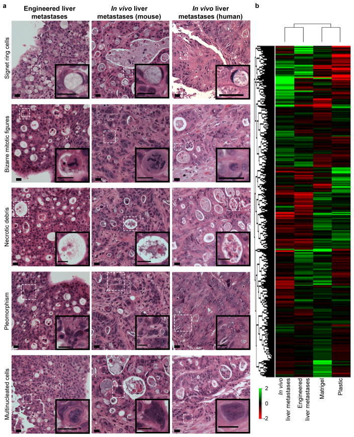
Figure 3.
Engineered liver metastases are comparable to liver metastases found in vivo. (a) Engineered HT-29 liver metastases (left panel), liver metastases formed following intrasplenic injection of HT-29 cells (middle panel), and liver metastases biopsied from late stage human colorectal cancer patients (right panel) all demonstrate classic histologic features of liver metastases of gastrointestinal origin. Experiments were repeated four times independently with similar results. Scale bars, 20 μm. (b) Hierarchal cluster analysis of average global gene expression patterns displayed by HT-29 cells grown on plastic (“Plastic”), Matrigel (“Matrigel”), liver BMSs (“Engineered liver metastases”), and in vivo liver metastases derived from intrasplenic injection of HT-29 cells (“In vivo liver metastases”) (n = 4 biologically independent samples).

