Figure 1.
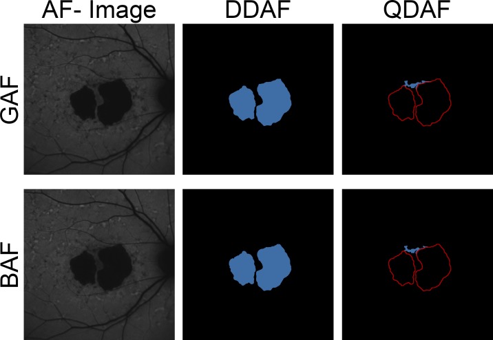
Grading of GAF and BAF images. Measurements of DDAF to QDAF decreased AF (≥90% darkness and 50%–90% darkness, respectively) were performed semiautomatically based on GAF (top) and BAF (bottom) images. In a first step, DDAF (blue area, middle) was annotated based on gray levels. Then, DDAF delineations were transferred to constraints (red lines, right) in order to measure QDAF (blue area, right). Despite absorption due to macular pigment, the high AF intensity in ABCA4-related retinopathy usually allows distinct demarcation and validation of foveal involvement.

