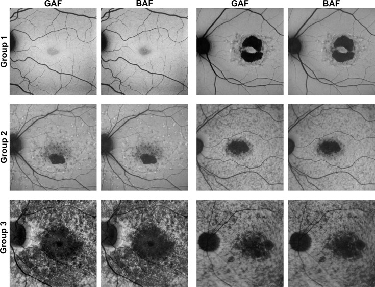
Figure 3.
Exemplary images of eyes separated by ERG-based classification. Two exemplary eyes of each ERG-based group38 are presented to demonstrate the high variability of ABCA4-related retinopathy and therefore different levels of interreader and intermodality agreement. The phenotypic spectrum of group 1 eyes ranges from eyes with only a central area of QDAF to eyes with DDAF surrounded by QDAF or flecks. Group 2 eyes typically revealed one or multiple well-circumscribed areas of DDAF with multiple hypo-and hyperautofluorescent flecks up to/over the vascular arcades. Group 3 usually contains eyes with multiple widespread hypoautofluorescent areas. Of note, images of both AF modalities differ only slightly, most obviously in the very mild phenotype of the first group 1 eye (top left).

