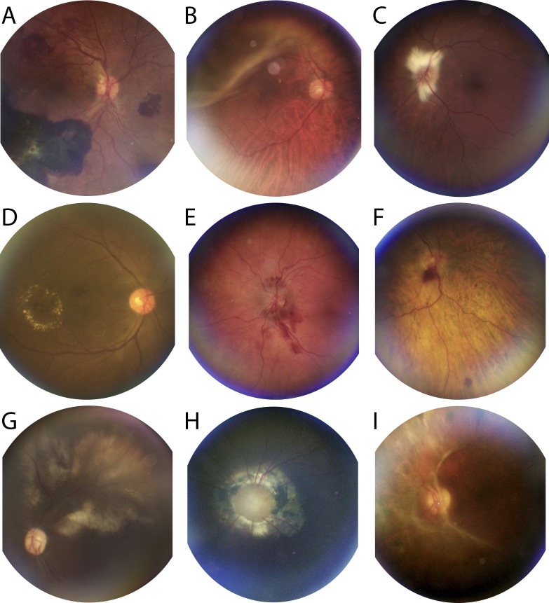
Figure 3.
CellScope Retina produces diagnostic-quality photographs of retinal abnormalities. (A) Hypertensive retinopathy with ruptured RAM along the inferior arcade with hemorrhage in multiple layers. (B) Superotemporal bullous macula-involving rhegmatogenous retinal detachment in the right eye extending from 9:00 to 1:00 o'clock. (C) Peripapillary myelinated nerve fiber layer with flame-shaped appearance and feathered borders in the left eye. (D) Circinate retinal exudates in the inferotemporal macula of the right eye. (E) Acute papilledema with flame hemorrhages extending from the optic disc and proximally along the inferior arcade of the left eye. (F) DR with dot-blot and disc hemorrhages in the right eye. (G) CMV retinitis demonstrating broad retinal whitening along the superior arcade of the left eye. (H) Morning Glory disc anomaly in the left eye. (I) Epiretinal membrane with fibrovascular changes in the left eye. All images were acquired after pharmacologic mydriasis.

