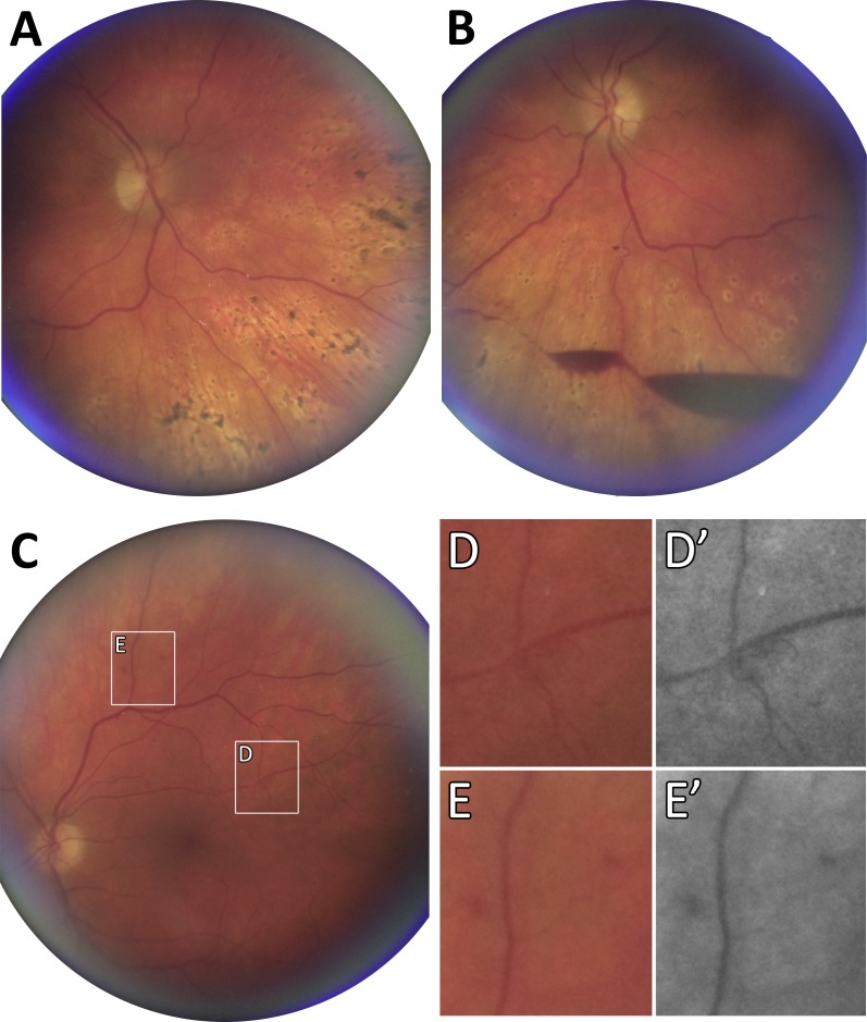
Figure 5.
Imaging of RWDR with CellScope Retina. Fundus photographs of a patient with diabetes mellitus with right (A) and left (B) eye demonstrating prior PRP and re-activation of quiescent diabetic retinopathy with preretinal hemorrhage in the left eye. (C) Fundus photograph of the left eye in a patient with diabetes mellitus but no known history of DR. CellScope Retina resolves trace signs of neovascularization (D) and sparse microaneurysms (E). Postprocessing with red-subtraction can be used to enhance contrast of vascular structures in the superficial retina, including neovascularization (D') and microaneurysms (E').

