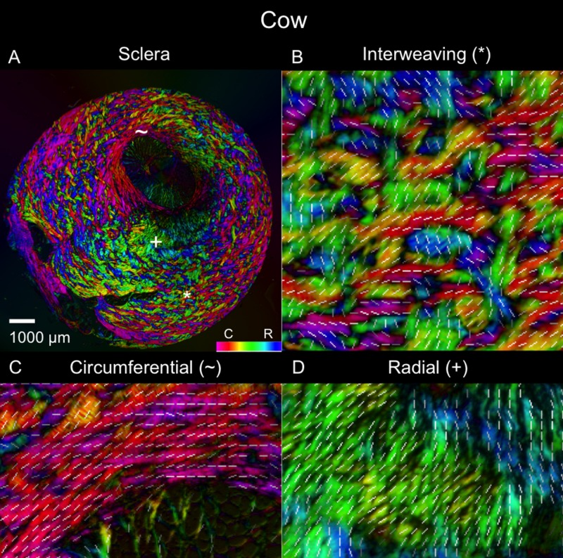
Figure 4.
Polar orientation of fibers in the cow eye. Shown are (A) an image of the whole section's energy-weighted polar orientation map and (B–D) close-ups of three regions: (B) interweaving fibers that form a basket-weave pattern (asterisk); (C) fibers oriented circumferentially (tilde symbol); and (D) fibers oriented radially from the canal (plus symbol). The colors represent the polar orientation or direction at each pixel, with the intensity scaled by “energy”, as described in the main text. In addition, to simplify discerning fiber orientation, we overlaid short white line segments representing the mean orientation over a small square region with side length equal to the length of the line.

