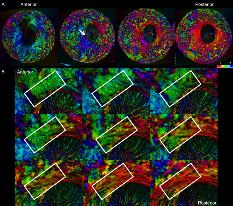
Figure 13.
Sequence of images of polar orientation in pig peripapillary sclera to illustrate the depth dependence of fiber orientations. (A) Four images of evenly spaced sections through depth, from anterior/innermost to posterior/outermost. Radial fibers are clearly discernible in the anterior-most section, especially from 5 to 10 o'clock. With increasing depth, radial fibers progressively give way to circumferential fibers, initially in the region immediately adjacent to the canal, pointed out by the white arrow. A wide ring of circumferential fibers is discernible in the deeper sclera, surrounded by regions of interweaving fibers of low anisotropy. (B) Images of a subset of consecutive sections through depth to visualize the transition from radial to circumferential fibers. The white boxes highlight the same region in each image. These areas of transition occurred most often between radial and circumferential regions.

