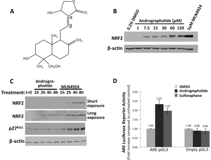
Fig 1. Andrographolide increases NRF2 protein expression and transcriptional activity.
(A) Structure of andrographolide. The α and β carbons of the Michael acceptor group are indicated. (B) HEK293T cells were treated with increasing andrographolide concentrations, as indicated, for 4 hours. DMSO was added as a negative control and the Cullin E3 RING ligase (CRL) inhibitor MLN4924 (1 μM) served as positive control. Cells were lysed and cell lysates analyzed by Western blotting using NRF2 and actin antibodies. (C) To measure the time dependence of andrographolide dependent NRF2 activation, HEK293T cells were treated with 7.5 μM andrographolide or 1 μM MLN4924 for the indicated times. Subsequently, cell lysates were analyzed by Western blotting with the indicated antibodies. The CRL substrate p27Kip1 served as a positive control for the CRL inhibitor MLN4924. (D) To measure Nrf2 transcriptional activity, the ARE luciferase reporter assay was performed as described under Materials and Methods. The cells were transfected for 24 hours before treatment with andrographolide (7.5 μM) or sulforaphane (10 μM) for 6 hours. The results shown are representative of two independent experiments.

