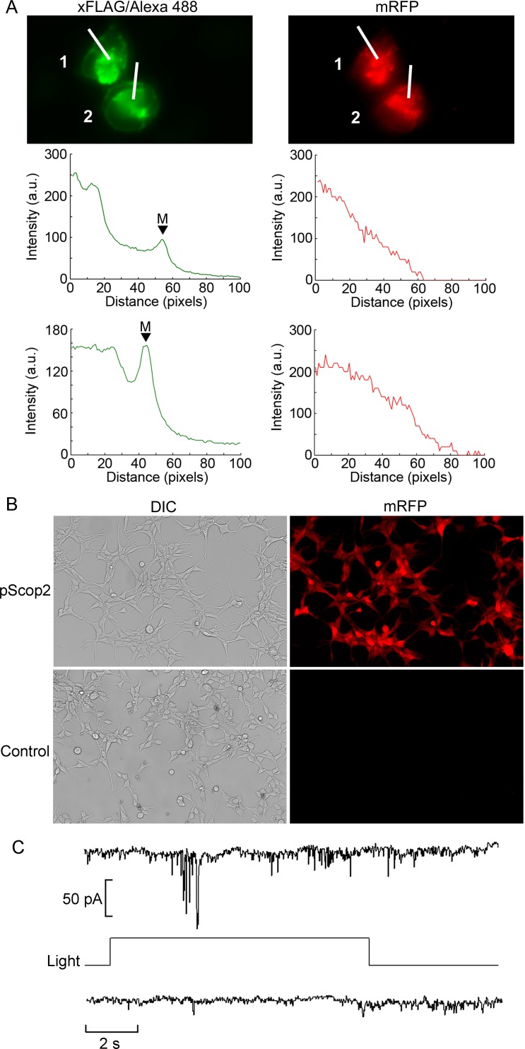
Fig 3. Functional expression of pScop2 in a HEK 293 cell line stably transfected with a bi-cistronic vector.
(A) Immunofluorescence of FLAG (left) and mRFP fluorescence (right) showing co-expression of the opsin and the reporter. Intensity profiles were obtained along the lines crossing the two cells, showing a different sub-cellular distribution in the two cases: pScop2 was abundant in the nuclear region, and also accumulated in the membrane (‘M’ & arrowheads), whereas mRFP was more distributed. (B) Low-magnification Nomarski and fluorescence micrographs of the reporter fluorescent protein of a transfected culture subjected to geneticin selection, and control cells. Percentage transfection in the former was 100%. (C) Recording of ion currents in the whole-cell modality, in a stably transfected HEK 293 cell pre-incubated with 11-cis-retinal to regenerate the photopigment. Presentation of a light step evoked a fluctuating inward current (top trace). In the absence of lightcurrent remained stable (bottom).

