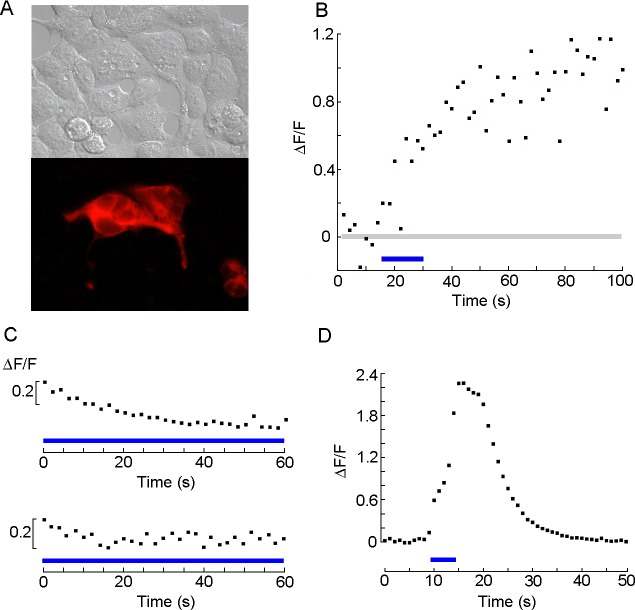
Fig 6. Functional expression of chimeric construct of pScop2 containing a Gq-specific ICL3.
(A) DIC and fluorescence microscopy of HEK-293 cells transfected with the bi-cistronic vector, showing a diffuse distribution of the mRFP. (B) Calcium fluorescence measurements in Fluo 4-loaded transfected cells. Brief pulses of epi-fluorescence illumination were delivered at 0.5 Hz, and fluorescence was repetitively monitored by a photomultiplier during those episodes; after 15 seconds, the same a light was applied in a sustained fashion (blue line) and subsequently the intermittent illumination protocol was resumed. The prolonged light induced a conspicuous increase in Ca fluorescence. (C) Control measurements in which Fluo-4 florescence was monitored in wild-type HEK293 cells incubated with 11-cis-retinal (top), or in HEK cells expressing pScop2-Gq but not exposed to the chromophore (bottom). A more prolonged irradiation with light, spanning the whole recording interval, was applied, and yet there was no indication of fluorescence increase.(D) Positive control, in which PLC-dependent Ca mobilization was activated by stimulating endogenous Gq-coupled muscarinic receptors with a saturating concentration of carbachol (20 μM). Fluorescence during the epi-illumination pulses was integrated.

