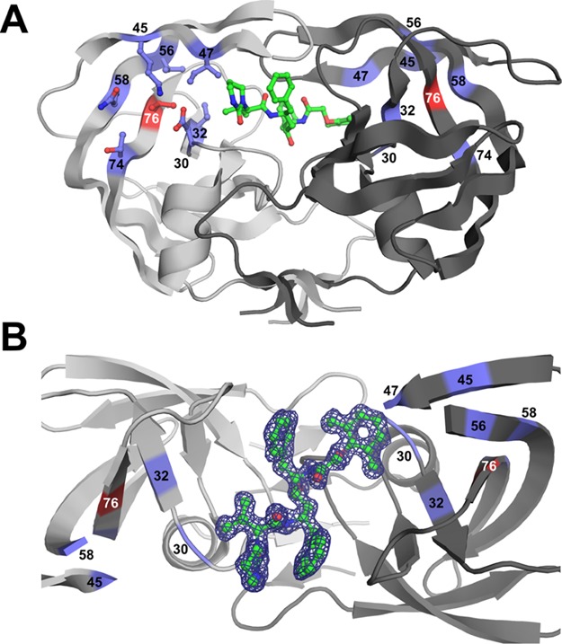
Figure 2.

Structure of the PRL76V dimer with inhibitor 2. (A) Structure of the PRL76V dimer in complex with inhibitor 2. The two subunits are shown in light and dark gray ribbons. Inhibitor 2 is shown with green bonds. The side chain of Val76 is in red sticks; side chains of residues that interact with the Leu76 side chain are shown as purple sticks. Side chains were omitted on second subunit for clarity. (B) Electron density map for single conformation of inhibitor 2 (green sticks) bound in the PRL76V dimer. 2Fo – Fc electron density map contoured at the 1.0σ level is represented by the purple mesh. The view is rotated about 90° from (A). Flap residues 46–57 in subunit (A) and 48–54 in subunit (B) have been removed for clarity.
