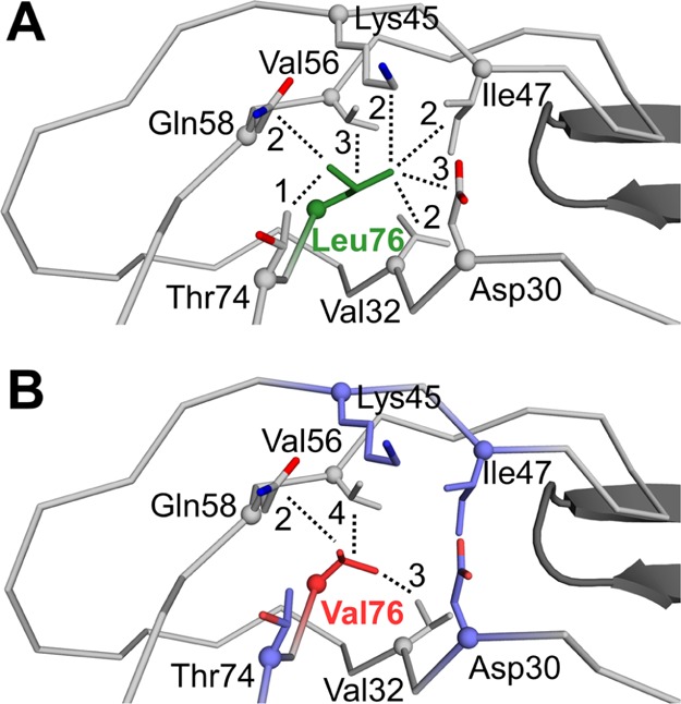
Figure 4.

Hydrophobic interactions of residue 76. Hydrophobic interactions are shown for inhibitor 3 complexes with (A) PRWT and (B) PRL76V. The main chain of PR is shown as gray ribbons. The side chains are represented by sticks with Cα atoms as spheres. The tip of the flap in the second subunit of the dimer is shown as black cartoons. Leu76 is green, Val76 is red, and side chains of residues that lose interactions in the mutant are shown in purple in (B). The number of van der Waals contacts of the side chain of residue 76 with neighboring residues is indicated next to each dotted line.
