. 2018 Sep 28;7:e39636. doi: 10.7554/eLife.39636

Figure 1. Single-cell RNA sequencing reveals molecularly distinct Pdgfrb-expressing subpopulations in visceral adipose tissue.

(A) Schematic overview of the MuralChaser model: a ‘Tet-On’ system allowing for indelible labeling of Pdgfrb-expressing cells. In the absence of doxycycline (Dox), gonadal SVF cells are labeled membrane tdTomato+ and are devoid of membrane GFP expression. In the presence of Dox, rtTA activates Cre expression in Pdgfrb-expressing cells. Cre excises the loxP-flanked membrane tdTomato (mtdTomato) cassette and allows constitutive activation of membrane GFP (mGFP) reporter expression. The gating strategy shows prospective isolation of tdTomato- GFP+ cells from the stromal vascular fraction of gonadal WAT (gWAT). (B) t-distributed stochastic neighbor embedding (tSNE) plot of 1045 tdTomato- GFP+ cells isolated from pooled gWAT depots from five male MuralChaser mice. Equal numbers of cells were combined from five individual mice for single-cell RNA-sequencing. Clustering was generated using k-means = 4. See Figure 1—source data 1. (C) Distribution of Gfp and tdTomato expression within tSNE plot. Transcript counts represent Log2 of gene expression. (D) Heatmap of top 20 most differentially expressed genes defining the clusters indicated in (B). See Figure 1—source data 1. (E) Gene expression distribution of adipocyte/adipogenesis-associated genes. (F) Gene expression distribution of genes associated with terminal adipocyte differentiation. (G) Gene expression distribution of genes associated with fibrosis and inflammation. (H) Gene expression distribution of mesothelial cell markers.

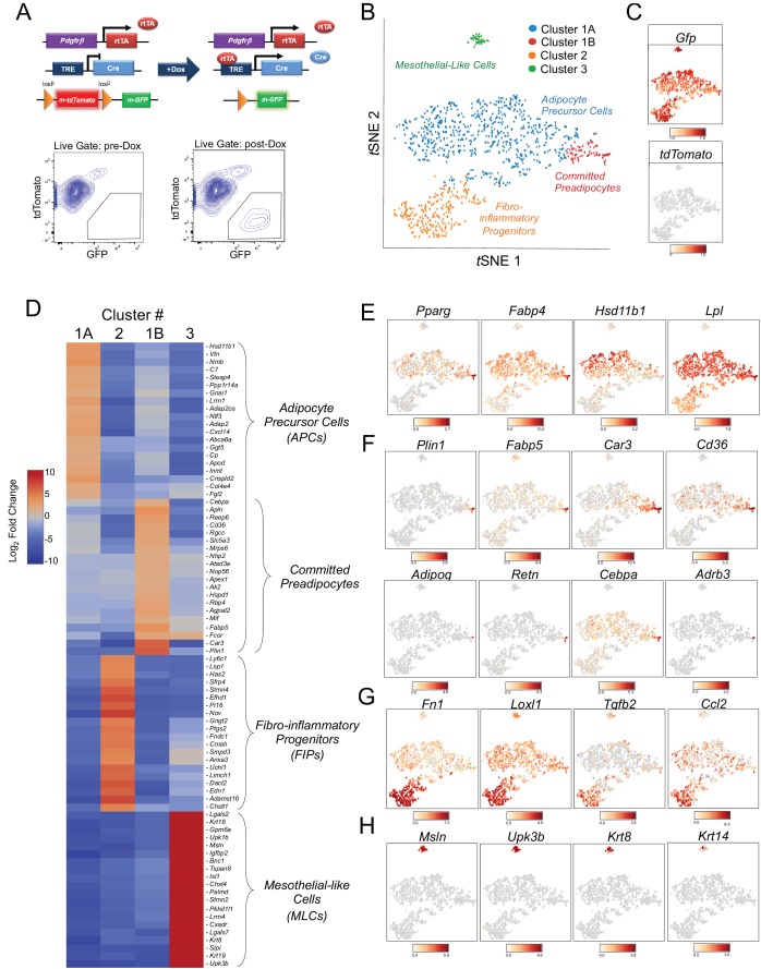
Figure 1—source data 1. Complete list of differentially expressed genes (k-means = 4).
DOI: 10.7554/eLife.39636.006

Figure 1.

Figure 1—figure supplement 1. GFP expression in gonadal WAT of MuralChaser mice.

Figure 1—figure supplement 1.

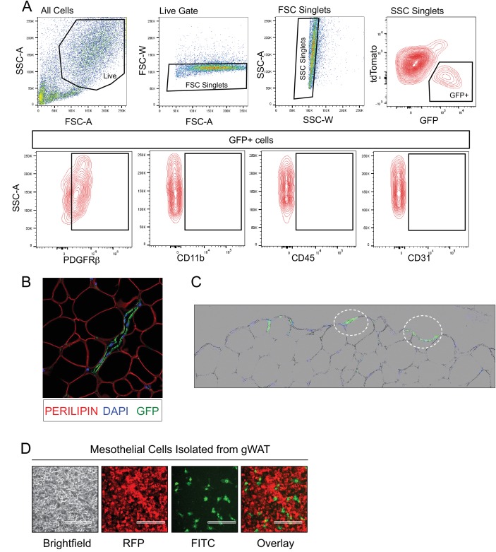
(A) Representative FACS gating strategy for the isolation of mGFP+ cells from gonadal WAT of MuralChaser mice and representative plots indicating the expression of PDGFRβ expression in these cells. mGFP+ cells from MuralChaser mice are devoid of CD31, CD45, and CD11b expression. (B) 63x confocal image of sectioned gonadal WAT obtained from doxycycline-treated MuralChaser mice. Paraffin sections were stained with antibodies raised against GFP and PERILIPIN, and counterstained with DAPI. Note the presence of GFP+ cells along the vasculature. (C) Digital overlay of 20x brightfield and fluorescent images of sectioned gonadal WAT obtained from doxycycline-treated MuralChaser mice. Paraffin sections were stained with antibodies raised against GFP and counterstained with DAPI. Note the presence of GFP+ epithelial like cells (circled) along the outer later of the depot where the mesothelium resides. (D) Fluorescent images of live cultures of mesothelial cells isolated from gonadal WAT from doxycycline-treated male MuralChaser mice. mGFP expression is found in a small subset of the cobblestone mesothelial-like cells within the cultures. Scale bar = 200 μm.
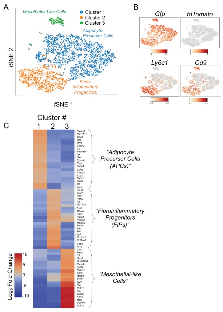
Figure 1—figure supplement 2. tSNE plot of 4203 tdTomato- GFP+ cells isolated from gonadal WAT of MuralChaser mice.

Figure 1—figure supplement 2.

(A) tSNE plot of 4203 tdTomato- GFP+ cells obtained from gonadal WAT of MuralChaser mice. (Median UMI count of 1873 per cell, mean reads per cell of 13,268, and median genes per cell of 908). (B) Distribution of Gfp, tdTomato, Ly6c1, and Cd9 expression within the identified clusters. (C) Heatmap of top 20 most differentially expressed genes defining the clusters indicated in (A).