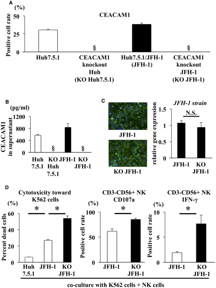
Figure 2.

Naïve NK cell cytotoxicity toward K562 cells was increased by coculture with CEACAM1 KO Huh7.5.1/JFH‐1 cells compared with Huh7.5.1/JFH‐1 cells. (A,B) CEACAM1 KO Huh7.5.1 cells were generated using the CRISPR/Cas9 system. Flow cytometry and ELISA were used to confirm that the CEACAM1 KO Huh7.5.1 cells and CEACAM1 KO Huh7.5.1/JFH‐1 cells did not express CEACAM1 at the cell surface and in supernatants, respectively. (C) Immunofluorescence staining for NS5A (green) and DAPI (blue) was performed in Huh7.5.1/JFH‐1 cells (left panel) and CEACAM1 KO Huh7.5.1/JFH‐1 cells (right panel). The efficiency of infection in Huh7.5.1/JFH‐1 cells was similar to CEACAM1 KO Huh7.5.1/JFH‐1 cells, as determined by qRT‐PCR (means ± SD, n = 4). (D) CFSE‐labeled K562 cells were added after 24 hours of NK cell coculture with Huh7.5.1 cells, Huh7.5.1/JFH‐1 cells, or CEACAM1 KO Huh7.5.1/JFH‐1 cells, and cocultures were incubated at 37°C. After 6 hours of incubation, 7‐AAD was added, and cytotoxicity was measured by flow cytometry (means ± SD, n = 4). NK cells were incubated with K562 cells and subjected to flow cytometry, to evaluate the expression of CD107a (means ± SD, n = 4) and the intracellular expression of IFN‐γ (means ± SD, n = 4). Representative data obtained from the peripheral NK cells of healthy subjects are shown. * P < 0.05. §Not detected. Abbreviation: N.S., not significant.
