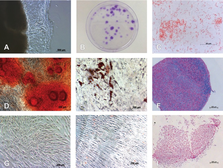
Figure 1.
Isolation and characterization of G-MSCs. (A) Microscopic appearance of outgrowing cells from free gingival margin connective tissue. (B) Microscopic appearance of CFUs of G-MSCs stained with crystal violet. (C) Gram-staining of Aggregatibacter actinomycetemcomitans (A.actinomycetemcomitans) colonies. (D) Alizarin Red staining of G-MSCs after osteogenic induction. (E) Oil Red O staining of G-MSCs after adipogenic stimulation. (F) Alcian Blue and nuclear-fast-red counter staining of G-MSCS after chondrogenic stimulation. (G) Alizarin Red staining of G-MSCs cultured in basic medium. (H) Oil Red O staining of G-MSCs cultured in basic medium. (I) Alcian Blue and nuclear-fast-red counter staining of G-MSCs cultured in basic medium.

